Radiofrequency neurotomy, also known as radiofrequency ablation (RFA), is a minimally invasive, outpatient procedure used to treat chronic back pain, neck pain, hip pain, and knee pain. The procedure uses controlled radiofrequency energy to disrupt specific sensory nerves responsible for transmitting pain signals.
Radiofrequency neurotomy is commonly performed after diagnostic nerve blocks confirm the source of pain.
What Is Radiofrequency Neurotomy?
Radiofrequency neurotomy uses heat generated by radio waves to create a small, precise lesion on a targeted nerve. This interrupts the nerve’s ability to transmit pain signals to the brain.
The procedure is most frequently used to treat:
Facet joint pain in the cervical and lumbar spine
Sacroiliac joint pain
Hip joint pain
Knee osteoarthritis pain
Pain relief typically develops within days to weeks and may last several months or longer, depending on nerve regeneration.
Conditions Treated
Radiofrequency neurotomy is indicated for chronic pain lasting longer than 12 weeks that has not improved with conservative treatment.
Common indications include:
Chronic low back pain due to facet joint arthritis
Chronic neck pain from cervical facet joints
Hip pain related to osteoarthritis
Knee pain from osteoarthritis
Sacroiliac joint pain
Spine pain following traumatic injury, including motor vehicle accidents
RFA is particularly effective when pain originates from arthritic or degenerative joints.
Candidate Criteria for Radiofrequency Ablation
You may be a candidate if you:
Have chronic back, neck, hip, or knee pain lasting more than 12 weeks
Have failed conservative treatments such as physical therapy and medications
Have experienced significant pain relief from diagnostic nerve blocks
Have imaging evidence of joint degeneration or injury
Diagnostic medial branch blocks or joint injections are typically performed prior to radiofrequency neurotomy to confirm the pain source.
Table of Contents
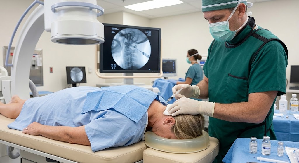
How the Procedure Is Performed
The patient is positioned comfortably in a procedure suite.
The skin is sterilized and locally anesthetized.
Using fluoroscopic (live X-ray) guidance, the physician advances a small needle into the atlanto-occipital or atlanto-axial joint.
Contrast dye may be used to confirm accurate placement.
Medication is injected into the joint space.
The procedure is performed on an outpatient basis and typically takes less than 30 minutes.
Benefits of Radiofrequency Neurotomy
Long-lasting pain relief compared to injections
Minimally invasive, non-surgical treatment
Reduced need for pain medications
Improved mobility and function
Outpatient procedure with minimal downtime
Radiofrequency Neurotomy (Radiofrequency Ablation) is available at our Jersey City, NJ office at 100 Town Square Place, Suite 405. Serving patients from Hoboken, Bayonne, Weehawken, and surrounding Hudson County communities. Learn more about pain management in Jersey City.
Begin your journey to recovery.
Reach out to our specialists to schedule an initial consultation. We’ll carefully review your symptoms and match you with the right expert for your specific condition.
CLINIC LOCATION
100 Town Square Place, Suite 405 Jersey City, NJ 07310