
Disclaimer: This article is intended solely for informational and educational purposes only. It does not constitute medical advice.
Pain from inside the hip joint is often felt deep in the groin; some people describe a “C-sign,” cupping the hand around the front and side of the hip. It may spread to the thigh or knee and worsen with walking, pivoting, deep squats, or prolonged sitting. Stiffness and pain with hip rotation can also occur. These clues suggest a hip source, but they do not identify one diagnosis by themselves.
What is FAI?
Femoroacetabular impingement (FAI) is contact between the hip socket and the femoral head–neck junction during movement. Cam morphology is extra bone or reduced roundness at the femoral head–neck junction. Pincer morphology is excess socket coverage, sometimes related to a deep or retroverted socket. Combined FAI includes both. These shapes are common in people without symptoms; FAI syndrome requires a matching pattern of symptoms, examination findings, and imaging.
What can a bedside exam tell you?
A clinician asks what activities reproduce pain, then checks gait, hip range of motion, strength, and function. The FADIR maneuver gently brings the hip into flexion, adduction, and internal rotation; reproduction of familiar groin pain supports suspicion of hip-joint irritation. FABER (flexion, abduction, external rotation) and measurement of internal rotation may add context. These tests help guide assessment, but a positive test alone cannot confirm FAI. Imaging and the overall clinical picture matter.
Why assess the spine and pelvis too?
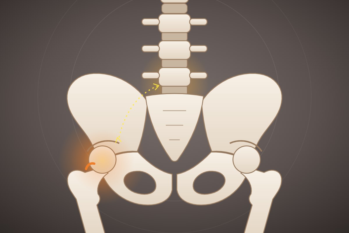
The hip, pelvis, and lumbar spine share movement and load. With FAI or hip osteoarthritis, reduced hip motion may change how the pelvis moves during sitting, standing, walking, or bending. Some people compensate with more lumbar movement; others have a stiffer spine or pelvis and rely more on hip motion. The pattern varies, so hip findings do not automatically explain back pain.
A clinician can observe forward bending and returning upright, single-leg stance, walking, and a squat, noting when the pelvis and low back begin to move relative to the hips. Comparing these tasks with hip range of motion helps describe lumbopelvic motion without assuming one “normal” pattern fits everyone.
Pain, control, and stability
Persistent pain involves more than tissue signals: the nervous system can change how it processes threat, sensation, and movement. Sensitivity may increase, but central sensitization is a specific neurophysiological term and cannot be diagnosed from pain duration alone. “Core” or pelvic instability usually describes a functional difficulty coordinating or tolerating load; it does not necessarily mean the bones or joints are structurally loose.
Pelvic function can be assessed with an active straight-leg raise: lying on the back, lift each straight leg and report effort or heaviness. A clinician may repeat it with gentle pelvic compression and compare symptoms and ease. Single-leg standing and walking provide additional information. These findings describe load transfer and symptoms, not a standalone diagnosis.
Weakness or a movement pattern?
True weakness is reduced force on resisted testing or dynamometry. A movement-system impairment is a repeatable pattern—such as trunk or pelvic compensation during a task—that changes symptoms or control. Clinicians compare isolated strength with repeated functional tasks and test whether cues or position changes alter movement. Both can coexist. This distinction helps tailor rehabilitation to the person’s capacity, coordination, and goals.