Disclaimer: This article is intended solely for informational and educational purposes only. It does not constitute medical advice.
Genicular nerve radiofrequency ablation, often shortened to GNRFA, is a minimally invasive treatment for chronic knee pain, especially pain related to osteoarthritis or persistent discomfort after knee replacement. Using imaging guidance, a specialist places needles near sensory nerves and applies radiofrequency energy to interrupt pain signals. The procedure does not remove the knee’s ability to move, rebuild cartilage, or cure arthritis; its goal is to reduce pain and improve function.
A recent International Pain and Spine Intervention Society (IPSIS) FactFinder asks whether GNRFA can cause osteonecrosis, sometimes called bone death. Osteonecrosis occurs when part of a bone loses enough blood supply that the tissue becomes damaged. Based on the published evidence available today, GNRFA has not been shown to increase the risk of osteonecrosis of the knee.
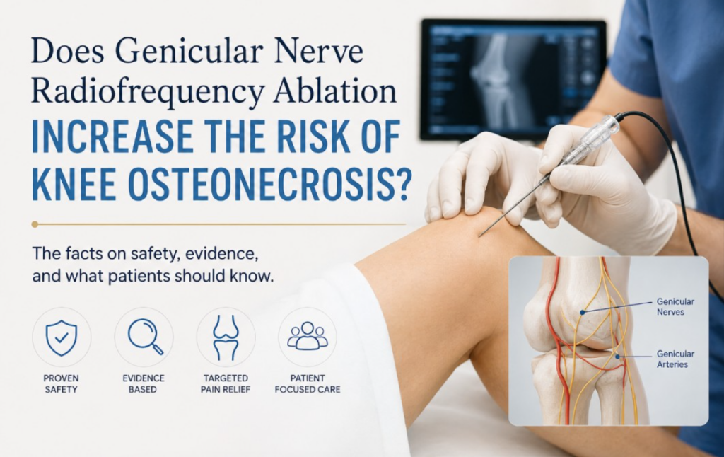
Why has the concern arisen? Genicular nerves travel close to genicular arteries around the knee. In theory, an unintended arterial injury could affect local circulation. A 2016 anatomical review identified vascular complications, including patellar osteonecrosis, in reports involving knee surgery and other procedures near these vessels. Importantly, those cases were not reports of osteonecrosis caused by GNRFA. At the time of that review, no vascular injuries following GNRFA had been reported.
Clinical outcomes also provide context. A real-world cohort followed 134 patients for an average of nearly two years; about half reported at least 50 percent pain reduction, and investigators described GNRFA as safe. Longer-term cooled RFA studies likewise reported sustained benefit without identified safety concerns. These findings cannot rule out exceptionally rare complications, but they offer no osteonecrosis signal.
The broader safety literature is reassuring but not absolute. Published GNRFA studies and reviews generally describe no complications or mostly temporary effects, such as soreness, bruising, swelling, or numbness. Rare but clinically important complications have included bleeding into or around the joint, infection, skin burn, and injury to the pes anserine tendon. Large, long-term studies designed specifically to detect very rare events remain limited, so “not demonstrated” should not be interpreted as “impossible.”
Technique matters. Careful patient selection, sterile preparation, appropriate imaging, and detailed knowledge of knee anatomy help clinicians avoid nearby blood vessels and other structures. Patients should tell their physician about blood thinners, bleeding disorders, infection, prior knee surgery, unusual anatomy, and medical conditions that may affect bone or circulation. A diagnostic genicular nerve block is often used to estimate whether interrupting these pain signals is likely to help before ablation is performed.
After GNRFA, follow the clinician’s recovery instructions and increase activity gradually. Contact the treating practice promptly for fever, expanding redness, drainage, severe swelling, new weakness, worsening pain, or inability to bear weight. These symptoms do not automatically indicate osteonecrosis, but they deserve medical evaluation.
The bottom line: current evidence does not establish that GNRFA raises the risk of knee osteonecrosis. The concern is anatomically plausible, yet published reports have not demonstrated a causal link. For appropriately selected patients, GNRFA remains a generally safe option that may provide meaningful pain relief. A personalized consultation can clarify expected benefits, alternatives, and individual risks.