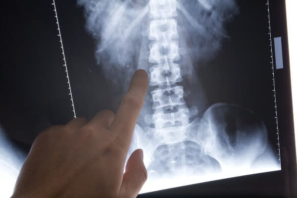
It is estimated that at least 15% of the US adult population suffer from chronic neck pain. While cervical intervertebral discs, cervical facet joints, atlanto-axial and atlanto-occipital joints, ligaments, fascia, muscle, and dura have all been shown to be potential causes for neck pain, it is often difficult to identify the exact cause of the pain. Spine imaging is often helpful in identifying targets for intervention.
Plain radiographs, MRI and CT scans have all been utilized as imaging modalities for diagnosing neck pain. Indications for radiographic imaging include age >50 with new symptoms, constitutional symptoms such as fevers or unexplained weight loss, moderate to severe pain lasting more than six weeks, progressive neurologic findings, infectious risk such as IVDA or immunosuppression, and history of malignancy.
Plain films often include a cervical series of seven views, including odontoid, lateral, AP, two oblique views, lateral view in flexion, and lateral view in extension. However, as the flexion and extension views have not been shown to lead to findings that changed clinical management, often they are excluded. Lateral views demonstrate vertebral alignment, screens for osteoarthritis and disc space narrowing, and may demonstrate bony pathology such as compression fracture. Oblique views are used to diagnose foraminal encroachment. AP views best show lateral spine deviation, while odontoid views are appropriate in acute trauma.
MRI and CT are more sensitive than plain films for diagnosing disc herniation, spinal cord compression, infection and malignancy. While MRI is preferred for soft tissue processes such as tumor, central stenosis, and disc herniation, CT is better for facet osteoarthrisis or other osseous changes. CT scans are not able to identify intramedullary pathology such as spinal cord tumors, and have the downside of significant radiation exposure.
In addition to spine imaging, fluroscopically guided diagnostic procedures such as cervical facet joint block have been shown to be helpful in establishing the etiology of neck pain. By injecting local anesthetic into the facet joint, the amount of immediate relief experienced by the patient is useful in determining if the facet joint is the source of pain. Cervical facet joint interventions such as therapeutic cervical medial branch blocks and radiofrequency neurotomy of medial branches in the cervical spine have fair evidence in terms of efficacy of pain relief. Intraarticular injections, on the other hand, had limited evidence for efficacy.
Fluroscopically guided epidural steroid injections have also been shown to be effective in providing short term relief of cervical radicular pain. Long term benefits are less certain, with few studies comparing the intervention to a true placebo group.
Spine imaging for chronic neck pain is helpful in both diagnosis and, when coupled with fluoroscopically guided pain procedures, provides therapeutic relief of this prevalent condition. Even general anesthesia providers should have a basic understanding of these concepts, as many of our patients are likely to have undergone similar evaluations and procedures.
References:
