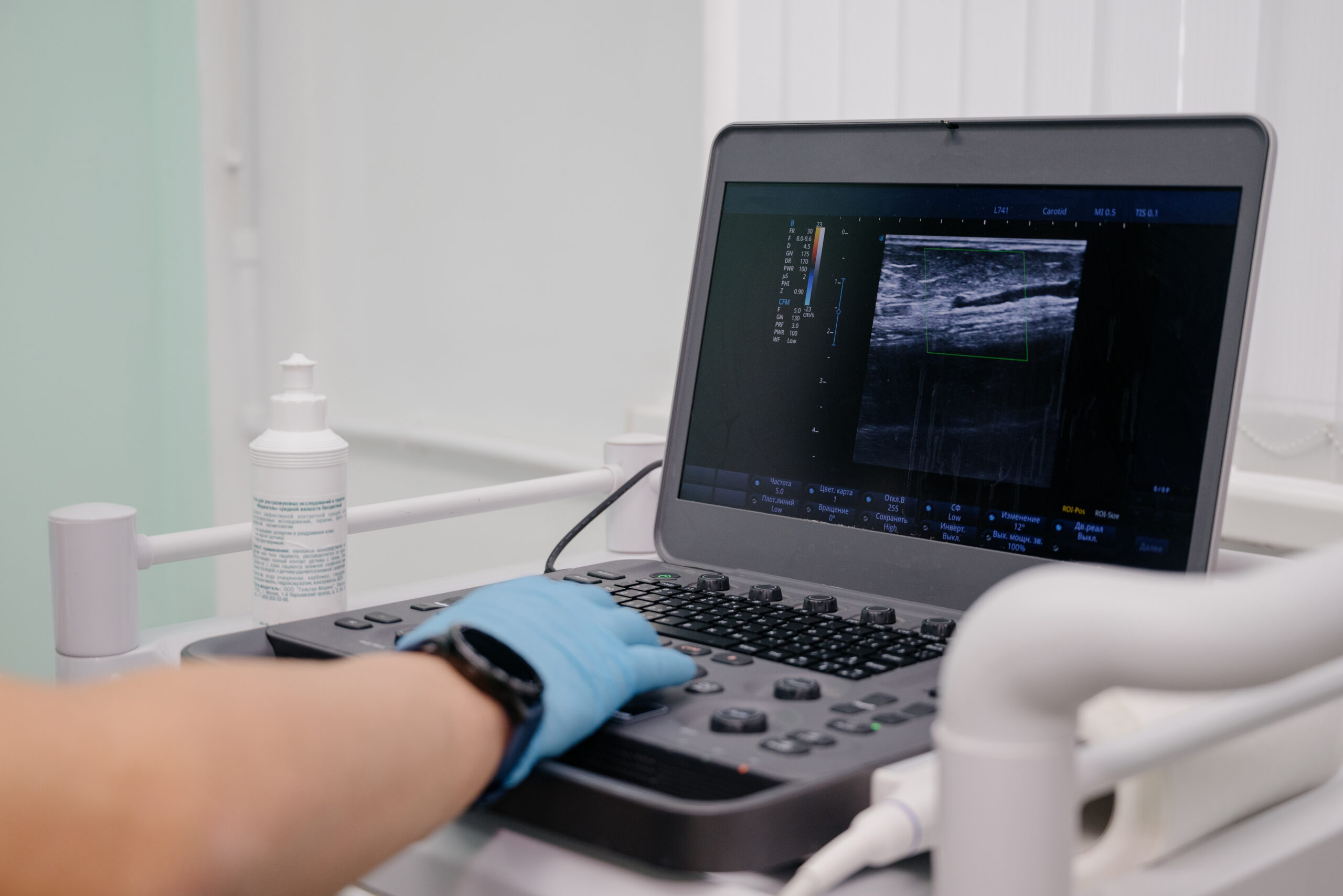
In the realm of interventional pain management, precision is paramount to ensure not only the efficacy of procedures but also the safety of patients. Ultrasound technology has revolutionized how medical professionals visualize vascular structures during various interventions, significantly reducing risks and improving outcomes. This article delves into the critical role of ultrasound in visualizing vascular structures during interventional pain management procedures, emphasizing its benefits and applications.
The Importance of Visualizing Vascular Structures
Accurately identifying and navigating around vascular structures during interventional pain management procedures is crucial to prevent complications such as vascular puncture or arterial damage. Such complications can lead to serious consequences, including bleeding, hematoma formation, and in severe cases, more significant vascular injuries that could require surgical intervention. Utilizing ultrasound allows clinicians to see real-time images of both the target treatment area and surrounding vascular anatomy, thereby enhancing procedural safety.
Advantages of Ultrasound over Traditional Methods
Traditionally, interventional procedures relied heavily on palpation and anatomical landmarks for guidance, which, while effective, come with limitations in accuracy and higher risks of vascular injury. Fluoroscopy, another common guidance technique, provides excellent real-time images of bony structures but falls short in visualizing soft tissues and vessels without the use of contrast agents. In contrast, ultrasound offers several distinct advantages:
- Real-time imaging: Ultrasound provides immediate feedback about the needle’s path and its relationship to surrounding tissues and vessels.
- Non-invasive and safe: This modality does not involve ionizing radiation, making it suitable for repeated use, particularly in vulnerable populations such as pregnant women or individuals with contraindications to radiation.
- Cost-effectiveness: Ultrasound machines are relatively portable and less expensive than other advanced imaging modalities, facilitating broader accessibility and utility in various clinical settings.
Applications of Ultrasound in Visualizing Vascular Structures
Ultrasound is invaluable in several key interventional pain management procedures where vascular visualization is crucial:
- Nerve Blocks: Whether performing a cervical plexus block or a sciatic nerve block, ultrasound guidance helps in avoiding vascular structures that are often in close proximity to nerve bundles.
- Joint Injections: Procedures such as hip or shoulder injections are high-risk due to the large vessels near the joint capsules. Ultrasound allows for precise placement of the needle, avoiding these vessels.
- Central Venous Access: Although not a pain management procedure per se, many patients undergoing long-term pain management may require central venous access for treatment administration. Ultrasound significantly reduces the risk of accidental arterial puncture or other vascular complications.
- Regenerative Medicine Injections: Techniques like platelet-rich plasma (PRP) or stem cell injections often target areas rich in vascular structures. Ultrasound ensures these biologics are accurately delivered, enhancing their efficacy and safety.
Best Practices for Using Ultrasound
To maximize the benefits of ultrasound in visualizing vascular structures during interventional procedures, adherence to best practices is essential:
- Proper Training: Clinicians should undergo thorough training in musculoskeletal ultrasound to adeptly identify vascular structures and understand the nuances of ultrasound-guided interventions.
- Pre-procedural Planning: A detailed ultrasound examination should be performed before the procedure to map out the vascular anatomy and plan the safest route for the intervention.
- Real-time Monitoring: Continuous ultrasound monitoring during the procedure allows for adjustments in real-time, enhancing safety and improving outcomes.
Conclusion
The visualization of vascular structures with ultrasound during interventional pain management procedures represents a significant advancement in medical technology, offering enhanced safety, efficacy, and patient satisfaction. As ultrasound technology continues to evolve, its applications in pain management are likely to expand, further embedding this essential tool in routine clinical practice. By providing clear, real-time images of vascular and other soft tissue structures, ultrasound not only improves the precision of interventions but also significantly mitigates the associated risks, marking a new era in the field of interventional pain management.
