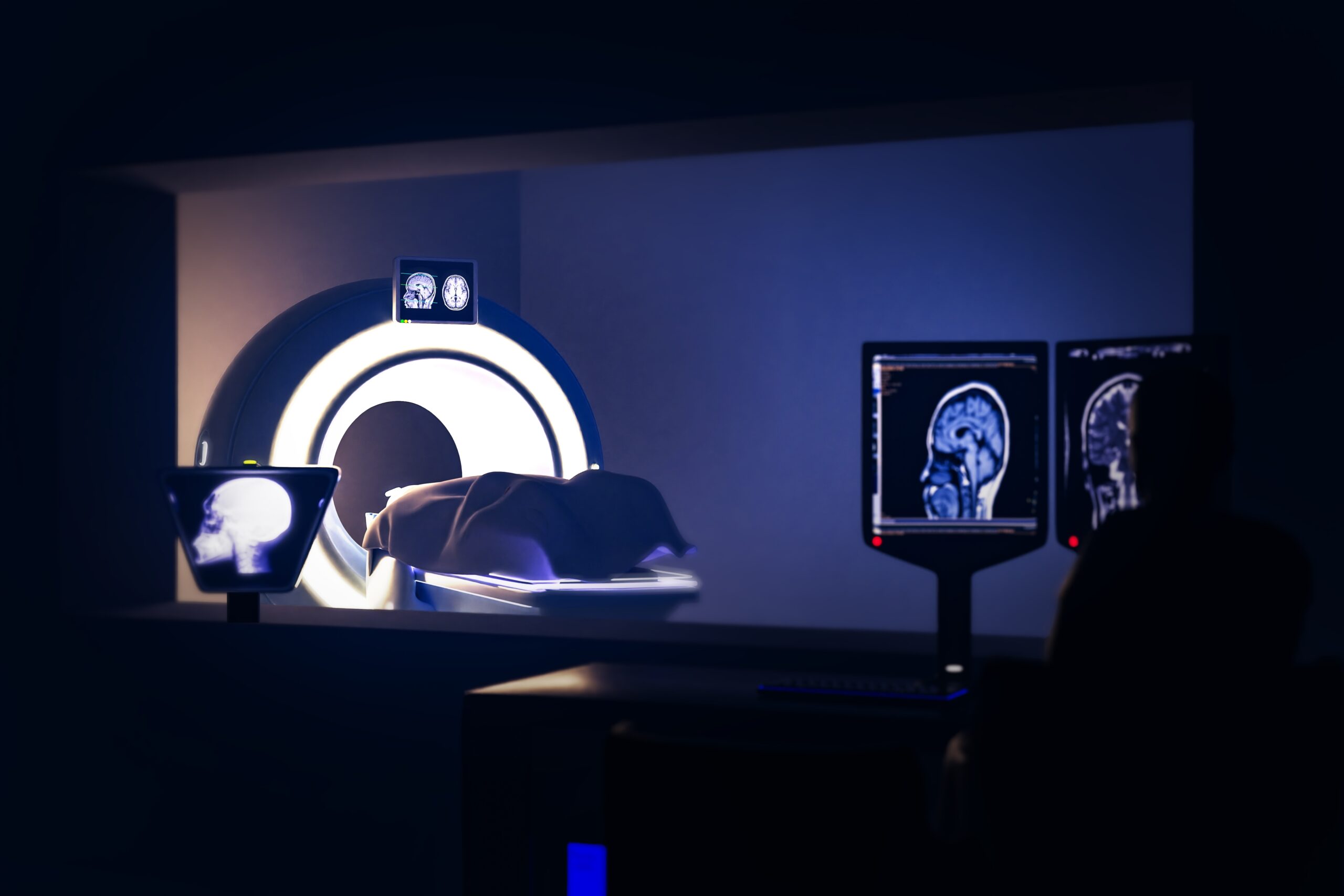
Magnetic Resonance Imaging (MRI) is a crucial diagnostic tool in modern healthcare, offering detailed images of internal body structures. However, for patients with spinal cord stimulation (SCS) devices, undergoing an MRI can present several barriers and challenges. This essay will discuss these barriers, including technical limitations, safety concerns, device interactions, and regulatory and procedural issues.
One of the primary barriers to MRI in patients with spinal cord stimulation is the technical limitations of the MRI machines themselves. MRI scanners use powerful magnets and radiofrequency (RF) waves to generate images. The strong magnetic field can interact with the metallic components of SCS devices, potentially causing movement or heating of the device. This interaction poses a risk of physical injury to the patient and can also damage the functioning of the SCS device. Moreover, the RF waves can induce electrical currents in the leads of the SCS system, leading to additional heating or unintended stimulation. These risks necessitate the use of MRI machines with specific settings or protocols when scanning patients with implanted devices, which may not be available in all medical facilities.
Safety concerns are another significant barrier. The interaction between the MRI’s magnetic field and the SCS device can lead to adverse events such as lead migration, tissue damage due to heating, or device malfunction. To mitigate these risks, stringent safety protocols must be followed. This includes thoroughly evaluating the MRI compatibility of the specific SCS device, as not all devices are designed to be MRI safe or conditional. Even in cases where devices are labeled as MRI conditional, they must be used within certain parameters of the MRI environment, such as specific magnetic field strengths and frequencies. Ensuring that these conditions are met can be challenging and time-consuming.
Moreover, the potential interactions between the MRI field and the SCS device can lead to diagnostic challenges. The presence of the device and its leads can cause artifacts in the MRI images, obscuring diagnostic information and reducing the quality of the images. This can be particularly problematic when imaging areas near the implanted device, such as the spinal cord or adjacent vertebrae. These artifacts may necessitate alternative imaging methods, which may be less effective or more invasive.
Regulatory and procedural issues also pose barriers. The compatibility of SCS devices with MRI is a relatively new development, and not all devices have been approved for use in this setting. This can limit the options for patients who require both SCS therapy and regular MRI scans. Additionally, the process of getting approval for an MRI scan in a patient with an SCS device can be cumbersome. It often involves coordination between multiple specialists, such as neurosurgeons, radiologists, and device manufacturers, to ensure that the procedure is conducted safely and effectively. This coordination can be time-consuming and may delay essential diagnostic imaging.
Finally, there is a need for increased awareness and education among healthcare providers about the challenges and risks associated with MRI in patients with spinal cord stimulation. Many clinicians may not be fully aware of the specific requirements and precautions needed to safely perform MRI scans in these patients. This lack of awareness can lead to delays in diagnosis or inappropriate use of MRI, putting patients at risk.
