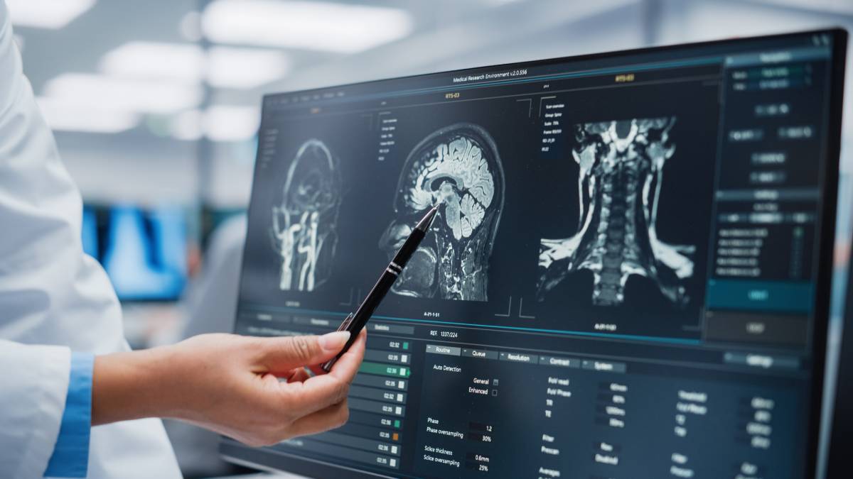
Medical imaging techniques were developed to visualize the structure and function of different tissues and organs in the body. In addition to X-ray radiography and X-ray endoscopy, other medical imaging techniques include computed tomography (CT), positron emission tomography (PET), magnetic resonance imaging (MRI), magnetic resonance spectroscopy (MRS), single-photon emission computed tomography (SPECT), digital mammography, diagnostic sonography, thermography, medical photography, electrical source imaging (ESI), tactile imaging, magnetic source imaging (MSI), medical optical imaging, and ultrasonic and electrical impedance tomography (EIT). The wide range of techniques creates many different uses for medical imaging.
X-ray medical imaging techniques utilize a beam of X-rays to visualize anatomical structure. Mammography is an application of X-ray medical imaging that uses an X-ray beam to generate images of breast tissue to visualize cancer cells. Digital mammography uses a computer-based system to produce superior images compared to those from film mammography. Angiography is another X-ray technique that visualizes blood vessels. CT scans use multidirectional X-Ray beams to generate a tomographic image. While X-ray images are 2D, CT images are 3D. CT scans produce superior images of tumors in soft tissues like the liver, brain, and lungs. PET & SPECT are types of CT scans differentiated by the mode of image production; the former uses radiopharmaceuticals to create three-dimensional images while the latter uses gamma rays.
MRI is widely used form of medical imaging that uses nonionizing radiation to generate images therefore has reduced risks compared to a CT scan. MRIs are known for their superior ability to visualize the skeleton and identify potential lesions in the bone tissue. Ultrasound medical imaging technologies use high frequency sound waves to visualize organs and tissues.1
Even with the many uses of current medical imaging, researchers continue seeking to improve the technology. Umirzakova et al. discuss the super resolution techniques underway to improve low resolution images commonly encountered in clinical practice. Super resolution techniques improve spatial resolution to create a more detailed imageby sidestepping intrinsic limitations of imaging. Techniques include Spatial, Transform, Hybrid, Traditional Machine Learning, and Deep Learning.
Interpolation is a conventional spatial technique that improves low resolution images but a newer technique – nonlocal interpolation draws on recurrent patterns in images to generate a more detailed real-time image. Hybrid methods combine spatial and transform techniques. Traditional machine learning extracts features from low resolution images to map high resolution images; the tool relies on the inherent similarities between the two groups such that it can map details to the high-resolution image. Tools which use traditional machine learning are Locally Linear Embedding (LLE) or Isomap. Deep learning utilizes a database of deep neural networks which are trained to recognize the similarities between low and high frequency images. The enhanced pattern recognition of deep learning allows the tool to extract and reconstruct features to develop a crisper more detailed image from a low resolution one.2
There are many beneficial uses of medical imaging, but at the same time, it should not be used excessively. For example, with X-ray-based techniques, providers must evaluate the risks associated with radiation exposure for individual patients. Overuse typically manifests through the referral process and is thought to be a feature of defensive medicine – a contemporary behavior among physicians which has the underlying focus of avoiding malpractice suits. One of the risks of medical imaging involves misdiagnoses – 10% of ultrasounds can show false positives for gallbladder stones and 67% can show thyroid nodules in asymptomatic patients. Another risk is cancer – an inherent aspect of radiation exposure.3
References:
- Hussain S, Mubeen I, Ullah N, Shah SSUD, Khan BA, Zahoor M, Ullah R, Khan FA, Sultan MA. Modern Diagnostic Imaging Technique Applications and Risk Factors in the Medical Field: A Review. Biomed Res Int. 2022 Jun 6;2022:5164970. doi: 10.1155/2022/5164970. PMID: 35707373; PMCID: PMC9192206.
- Sabina Umirzakova, Shabir Ahmad, Latif U. Khan, Taegkeun Whangbo, Medical image super-resolution for smart healthcare applications: A comprehensive survey, Information Fusion, Volume 103, 2024, 102075, ISSN 1566-2535, https://doi.org/10.1016/j.inffus.2023.102075 (https://www.sciencedirect.com/science/article/pii/S1566253523003913)
- Salerno, S., Laghi, A., Cantone, MC. et al. Overdiagnosis and overimaging: an ethical issue for radiological protection. Radiol med 124, 714–720 (2019). https://doi.org/10.1007/s11547-019-01029-5
