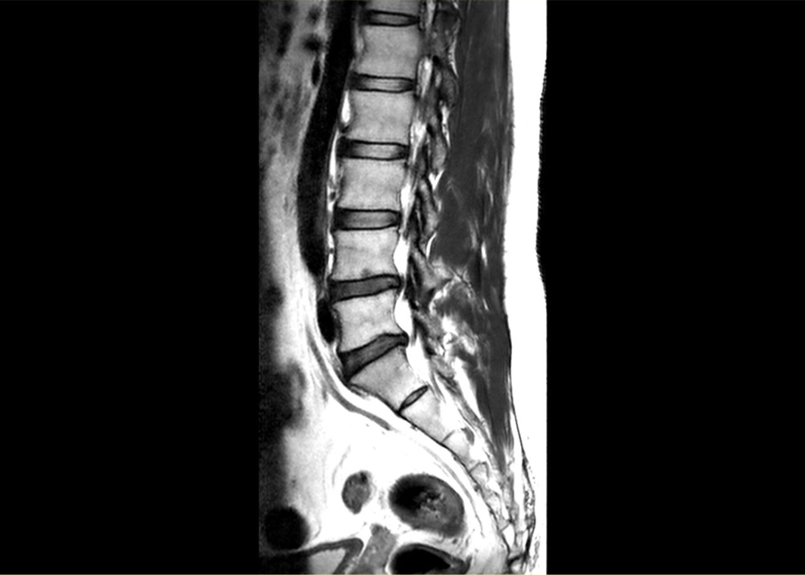
The human spine, a complex structure interwoven with a network of bones, nerves, muscles, and ligaments, is crucial for the upright posture and mobility that distinguishes Homo sapiens from other species. It also encases the delicate spinal cord, our body’s principal communication highway. Given its critical nature, any disorder or disruption can lead to significant pain, disability, or even paralysis. Diagnosing and treating issues within this intricate structure, therefore, demands a blend of precision and minimal invasiveness. This is where Spinal Canal Endoscopy comes into play.
Understanding Spinal Canal Endoscopy
Spinal Canal Endoscopy, often simply referred to as spinal endoscopy, is a procedure that employs advanced optics and miniaturized instruments to visualize, diagnose, and treat various spinal conditions. Unlike traditional open surgeries, which require significant incisions and extensive tissue disruption, spinal endoscopy allows surgeons to access the spinal canal through tiny incisions, minimizing collateral damage to surrounding tissues.
The procedure involves inserting a thin, flexible tube called an endoscope, equipped with a light source and a camera, into the spinal canal. This provides real-time, high-definition visuals of the internal anatomy, enabling the surgeon to navigate the tight confines of the spine with unmatched precision.
Applications and Advantages
- Diagnosis: Traditionally, spinal conditions were diagnosed using imaging modalities like X-rays, CT scans, or MRIs. While they provide a wealth of information, they can’t always capture the nuanced abnormalities or inflammatory processes at play. Spinal endoscopy offers direct visualization of pathological sites, allowing for a more accurate diagnosis.Therapeutic Interventions: Beyond mere visualization, spinal endoscopy is employed for a variety of treatments. Herniated discs, spinal stenosis, tumors, and some forms of infections can be addressed using endoscopic techniques. Specialized tools can be introduced through the endoscope to remove or repair damaged tissue, decompress nerves, or extract pathological masses.Minimal Invasiveness: A primary advantage of spinal endoscopy is its minimal invasiveness. This translates to reduced hospital stays, faster recovery times, lesser post-operative pain, and minimal scarring. For patients, this means a quicker return to normal activities and a reduction in overall healthcare costs.Preservation of Spinal Anatomy: Traditional open spine surgeries may necessitate the removal or disruption of significant amounts of bone or soft tissue. Endoscopic techniques, on the other hand, prioritize the preservation of the spine’s natural anatomy, leading to better long-term outcomes and reduced chances of complications.Safety: While every surgical procedure carries inherent risks, the direct visualization afforded by spinal endoscopy can reduce the chances of accidental nerve damage or other complications.
Challenges and Considerations
However, spinal endoscopy is not without its challenges. The learning curve for surgeons is steep, given the demand for exceptionally fine motor skills and spatial awareness. Proper training and experience are essential to achieve consistent and positive outcomes.
Patient selection is also crucial. Not every spinal condition is amenable to endoscopic intervention. Comprehensive evaluation, encompassing clinical examinations and traditional imaging studies, is necessary to determine if a patient is a suitable candidate.
Conclusion
The continued evolution of medical technology has provided us with tools that once seemed the stuff of science fiction. Spinal Canal Endoscopy stands as a testament to this progression, offering a fusion of diagnostic clarity and therapeutic finesse. While it may not replace traditional methods, it undeniably widens the spectrum of options available to both clinicians and patients, edging us closer to a future where spinal ailments can be addressed with maximal efficacy and minimal disruption.
