Ultrasound technology has revolutionized medical imaging, offering a non-invasive, real-time, and relatively affordable method to visualize internal structures. One of the specialized applications of ultrasound is in musculoskeletal imaging, where it plays a crucial role in diagnosing and managing conditions related to muscles, tendons, ligaments, nerves, and joints. Central to the effectiveness of musculoskeletal ultrasound is the transducer, or probe, used to emit and receive sound waves. Understanding how ultrasound transducers differ for musculoskeletal imaging involves exploring their design, frequency ranges, and specific features that enhance their utility for this application.
Design and Shape of Transducers
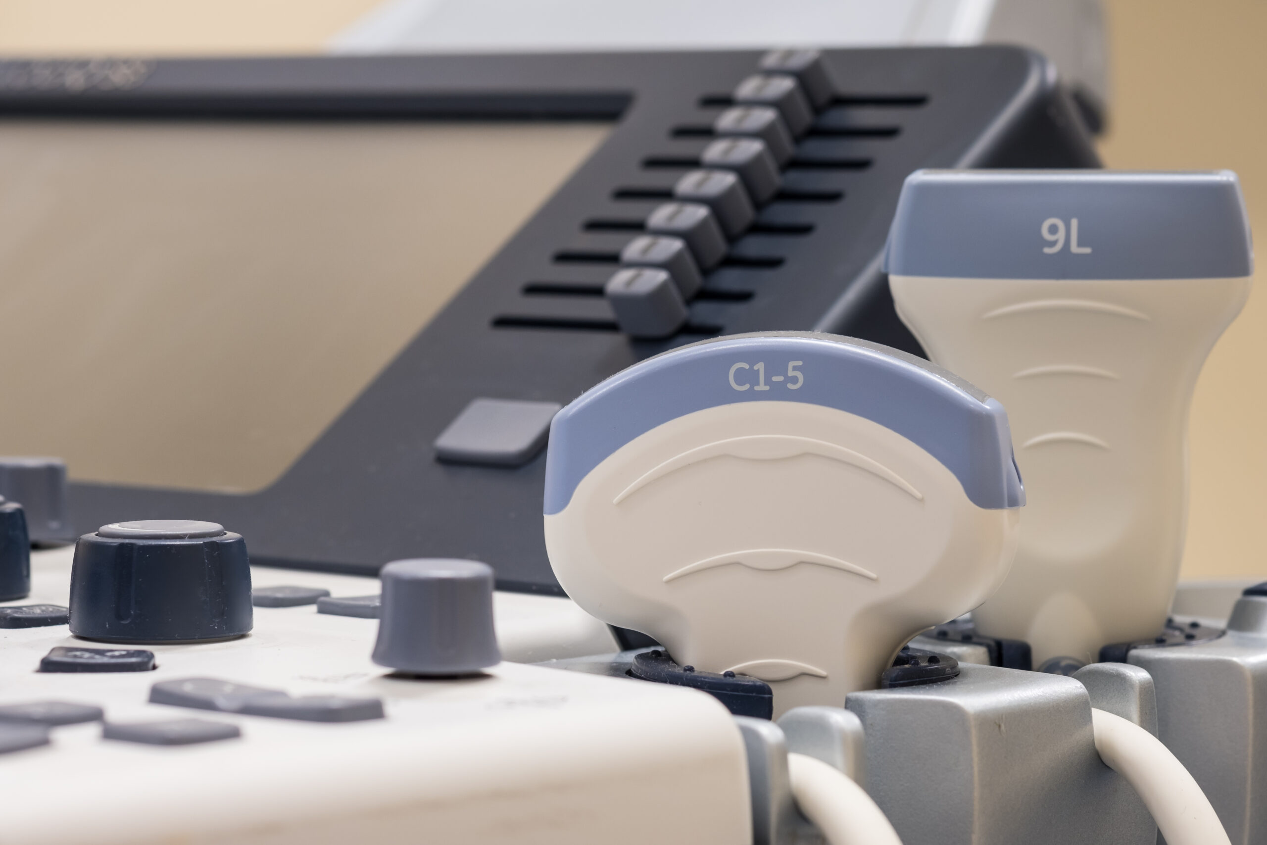
The design and shape of ultrasound transducers are critical in determining their suitability for various imaging tasks. For musculoskeletal imaging, transducers are typically linear in shape. Linear transducers have a flat surface, which allows for better contact with the skin and produces high-resolution images. This flat surface is particularly beneficial for imaging superficial structures like tendons and ligaments that are close to the skin surface.
In contrast, other types of ultrasound applications might use convex or phased array transducers. Convex transducers, with their curved surface, are more suitable for imaging deeper structures, such as in abdominal scans. Phased array transducers are used in cardiology due to their ability to image between the ribs and visualize the heart. Therefore, the flat, linear design of musculoskeletal transducers is a distinct feature tailored to the specific needs of visualizing musculoskeletal structures.
Frequency Range
The frequency range of an ultrasound transducer is one of its most defining characteristics. For musculoskeletal imaging, high-frequency transducers are preferred, typically ranging from 7.5 MHz to 15 MHz or higher. High-frequency transducers provide better resolution images, which is essential for visualizing the fine details of musculoskeletal tissues. Higher frequencies have shorter wavelengths, which improve the resolution of the images, allowing clinicians to see small structures and subtle differences in tissue texture.
However, high-frequency sound waves have limited penetration depth, making them less suitable for imaging deeper structures. This limitation is generally not an issue in musculoskeletal imaging, as most of the structures of interest, such as tendons, muscles, and superficial nerves, are located near the skin’s surface. Lower frequency transducers, which penetrate deeper but provide lower resolution images, are more suitable for abdominal or obstetric imaging.
Specific Features and Technology
Musculoskeletal ultrasound transducers incorporate several specific features and technologies to enhance their imaging capabilities. One such feature is the use of multiple focal zones. By using multiple focal zones, the transducer can focus on different depths within the tissue, improving the overall image clarity. This feature is particularly useful in musculoskeletal imaging, where different structures may lie at varying depths.
Another important feature is the use of harmonic imaging. Harmonic imaging enhances the quality of ultrasound images by using the harmonic frequencies generated by the tissues rather than the fundamental frequency emitted by the transducer. This technique reduces noise and artifacts, resulting in clearer images. This is especially beneficial in musculoskeletal imaging, where precise visualization of structures like tendons and ligaments is crucial for accurate diagnosis.
Additionally, musculoskeletal transducers may incorporate advanced Doppler capabilities. Doppler ultrasound is used to assess blood flow, which can be important in evaluating conditions such as tendinopathy or muscle tears, where altered blood flow patterns may be present. High-resolution power Doppler or color Doppler imaging can help identify increased vascularity associated with inflammation or injury.
Ergonomics and User Interface
The ergonomic design of musculoskeletal transducers is another critical aspect that sets them apart. These transducers are often designed to be lightweight and easy to handle, with a focus on reducing user fatigue during prolonged use. The design typically includes features such as a comfortable grip and easy access to control buttons, allowing the operator to adjust settings without losing focus on the imaging task.
The user interface of ultrasound machines used for musculoskeletal imaging is also optimized for this application. These systems often come with preset protocols and settings tailored for musculoskeletal examinations. This includes specific imaging modes, measurement tools, and reporting templates designed to streamline the workflow and improve diagnostic accuracy.
Clinical Applications and Benefits
The specialized design of musculoskeletal ultrasound transducers offers numerous clinical benefits. High-resolution imaging enables detailed assessment of small structures like tendons, ligaments, and nerves, facilitating the diagnosis of conditions such as tendonitis, ligament tears, and nerve entrapments. The real-time imaging capability allows for dynamic assessment of these structures, providing valuable information about their function and response to movement.
Ultrasound-guided interventions are another significant advantage of musculoskeletal transducers. Procedures such as injections, aspirations, and biopsies can be performed with greater accuracy and safety under ultrasound guidance. The ability to visualize the needle in real time ensures precise delivery of therapeutic agents or accurate sampling of tissues.
In sports medicine, ultrasound is widely used for the assessment and management of injuries. The portability of ultrasound machines equipped with musculoskeletal transducers allows for on-site evaluation of athletes, facilitating prompt diagnosis and treatment. This can be crucial in preventing further injury and optimizing recovery.
Conclusion
Ultrasound transducers designed for musculoskeletal imaging are distinct in their linear design, high-frequency range, and specialized features that enhance image quality and diagnostic accuracy. The ergonomic considerations and optimized user interfaces further support their effective use in clinical practice. These transducers play a vital role in the detailed assessment of musculoskeletal structures, enabling accurate diagnosis, guiding interventions, and supporting the management of a wide range of musculoskeletal conditions. As ultrasound technology continues to advance, the capabilities of musculoskeletal transducers are likely to expand, offering even greater benefits to clinicians and patients alike.