Pain is one of the most universal sensations that all humans experience at one time or another during their lifetimes. Most common is acute pain, which lasts for a short period of time, whereas chronic pain is an unresolvable pain that lasts for three or more months [7]. Accurate measures of pain are critical for treatment, research, and diagnosis.
Acute pain is usually in an isolated location, occurs as a result of an injury or surgical procedure, and can be successfully treated with mild medications, environmental changes, and stress reduction [7].
Chronic pain can occur when sensory pathways continue to transmit the feeling of pain even after the underlying condition has healed, and may require opioids or sedatives to manage [7]. Chronic pain affects 1 in 5 adults and can be a symptom of certain other diseases such as cancer or HIV/AIDS [7]. According to the National Health Interview Survey, in 2012, over 11% of the adult population the United States reported feeling pain daily [6]. In addition, the national cost for pain ranges from $560 to $625 billion dollars annually between healthcare costs, treatments for pain, and lost productivity [6].
For such a common experience, however, it is also one of the most difficult feelings to quantify or accurately measure. Accurate measurement is crucial as pain is an important clinical symptom that can be used to detect the degree of an injury, and better pain prediction will lead to better treatment [2]. Currently, however, the most common form of pain assessment is still self-reporting, in which the patients are asked to assess their own pain based on certain numerical, visual, or verbal rating scales [4][5]. Certain patient groups, such as patients with communicative impairments or disturbances of consciousness, older patients with dementia, or young children, may be unable to independently communicate their own pain [2]. Especially for these patient groups, objective, methods for physiological measures of pain should be designed to measure pain intensity sensitively and specifically [5].
There are many biomarkers that vary in the presence of pain. The goal of current research is to find one that is not only accurate and reproducible, but easy to sample and quick to measure [5]. Some examples of biomarkers that may indicate pain include changes in heart rate, respiratory rate, blood pressure, oxygen saturation, and hormonal changes [3]. Although these factors are known to correlate with pain, no test has been identified that can directly and accurately map these changes to a pain intensity scale.
One of the most promising methods so far is Magnetic Resonance Imaging (MRI) scans, which is able to measure both acute and chronic pain and, along with Positron Emission Tomography (PET) and Near-Infrared Spectroscopy, does so by assessing how activity in the spinal cord and brain changes depending on the quality, location, and duration of painful stimuli [1][5].
Another objective measure relies on cardiovascular and respiratory parameters, as heart rate variability, patterns of blood pressure and heart rate response [5]. The presence of noxious stimuli and interactions between the sympathetic and parasympathetic nervous systems can both cause changes to the time and frequency of intervals between consecutive heartbeats [5][7]. Since noxious stimuli induces minor increases in blood pressure and heart rate, its presence can be quantified using a sphygmomanometer and given a score out of 100 on the Cardiovascular Depth of Analgesia Index [5].
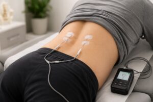
Two more biomarkers that have been linked with pain are skin conductance and pupillary changes. Sweating is a natural consequence of the autonomic nervous system being stimulated by noxious stimuli; thus, measuring the amplitude and frequency of skin conductance with adhesive electrodes can indicate the presence and intensity of pain [5]. Unfortunately, skin conductance responses to pain have only been consistently found in healthy adults, and more research on different patient groups is needed to further investigate this correlation [1]. Next, using infrared video pupillometry to measure changes in pupillary diameter and the light-induced pupillary dilation reflex can also indicate the presence of noxious stimuli and acute pain [1][5]. The challenge in pupillometry is that external factors such as drugs, environmental light, and patient age all can affect a patient’s pupillary as well, and thus the changes cannot be solely credited to pain [5].
As of today, many physiological indicators of pain have been identified, but none are reliable enough to be depended upon alone without confirmation by self-reporting [3]. Continued research is needed to develop objective, physiological measures of pain for high quality patient care.
References
[1] Korving, H. (2020). Physiological Measures of Acute and Chronic Pain within Different Subject Groups: A Systematic Review. Hindawi. https://doi.org/10.1155/2020/9249465
[2] Chu, Y., Zhao, X., Han, J., & Su, Y. (2017). Physiological Signal-Based Method for Measurement of Pain Intensity. Frontiers in Neuroscience, 11. https://doi.org/10.3389/fnins.2017.00279
[3] The Royal Children’s Hospital Melbourne. https://www.rch.org.au/rchcpg/hospital_clinical_guideline_index/Pain_assessment_and_measurement/
[4] Pain Assessment. (2020, February 4). Physiopedia. https://www.physio-pedia.com/index.php?title=Pain_Assessment&oldid=229642
[5] Cowen, R., Stasiowska, M. K., Laycock, H., & Bantel, C. (2015). Assessing pain objectively: the use of physiological markers. Anaesthesia, 70(7), 828–847. https://doi.org/10.1111/anae.13018
[6] Weir, K. (2017, November). Researchers are closing in on objective ways to measure pain. Monitor on Psychology. https://www.apa.org/monitor/2017/11/measure-pain
[7] Robertson, L., & Schmid, S. W. ATrain Education. 3. The Physiology of Pain | ATrain Education. https://www.atrainceu.com/content/3-physiology-pain